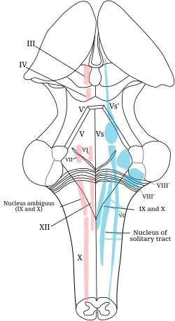
Cranial Nerve Nuclei Present In Floor Of 4th Ventricle
Cranial Nerve Nuclei Anatomy And Embryology Kenhub

Brainstem Ii Pons And Cerebellum Part 2 Cranial Nerves Skeletal System Anatomy Spinothalamic Tract

Abducens Vi Cranial Nerves Craniosacral Therapy Cranial Nerves Abducens Nerve

Mesencephalic Nucleus Google Search Cranial Nerves Pharmacology Nursing Cranial Nerves Mnemonic

Brainstem I The Medulla Organization Of The Central Nervous System Part 2 Nervous System Parts Medical Knowledge Nervous System

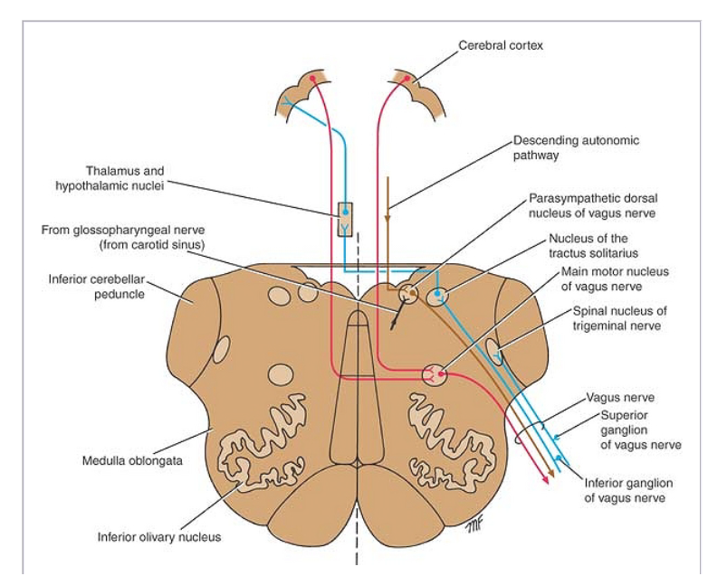
Main Motor Nucleus Nucleus Ambiguus Deep In The Reticular Formation Parasympathetic Nucleus Dorsal Nucleu Nervous System Parts Vagus Nerve Cranial Nerves
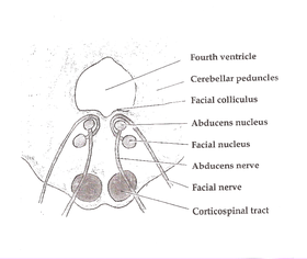
Multiple cranial nerve nuclei are located on the floor of the fourth ventricle with a high risk of permanent damage.
Cranial nerve nuclei present in floor of 4th ventricle.

Frontal Section Taken Through The Level Of The Rostral Diencephalon Where The Thala Mus Is Not Present Note Again Th Gross Anatomy Brain Anatomy Brain Parts
Hypoglossal Nucleus Wikipedia
Snp Cranial Nerve Nuclei Fa 2019 Kaplan 2018 Deja Review Hy 5th Edi Snell Flashcards Memorang

Jaypeedigital Ebook Reader
Source : pinterest.com
